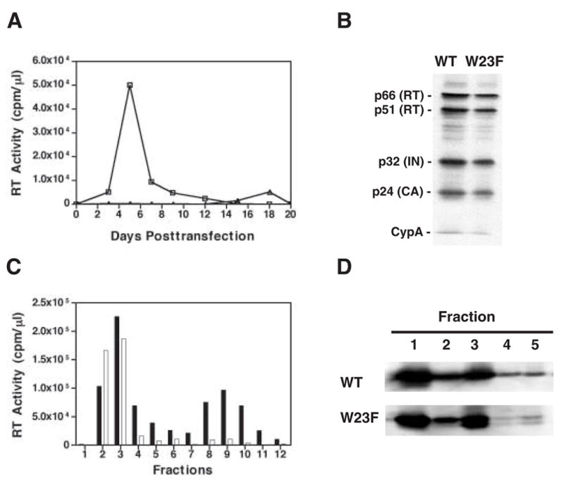
Fig. 2.

W23F phenotype. (A) Replication kinetics. MT-4 cells were transfected with WT and W23F plasmid DNAs. Cells were split 1:3 every 2 or 3 days. Virus production was monitored by assaying RT activity in the culture supernatants for each time point. Symbols: WT, open squares; W23F, open triangles; mock, open circles. (B) Incorporation of CypA into mature virus particles. HeLa cells were transfected with WT and W23F plasmid DNAs. Virion-associated proteins as well as CypA were detected by Western blot analysis, using anti-HIV RT and IN (Klutch et al., 1998) as well as anti-CA and anti-CypA (Tang et al., 2003b). The positions of individual viral proteins and CypA are indicated to the left of the gel. (C) Retention of HIV-1 RT in WT and W23F viral cores. Virions isolated from the supernatant fluids of transfected HeLa cells were treated with 0.3% NP-40 and were sedimented through 20% to 70% (wt/wt) linear sucrose gradients at 4°C for 16 h at 120,000 × g in a Beckman SW55Ti rotor (Tang et al., 2003b). Twelve fractions were collected from the top of the gradient. The bar graph shows RT activity in the fractions collected from WT (solid bars) or W23F (open bars) samples. (D) Retention of HIV-1 CA protein in viral cores. Detergent-treated virions were sedimented through sucrose step gradients centrifuged at 4°C for 60 min at 120,000 × g in a Beckman SW55Ti rotor (Tang et al., 2003b). Five fractions were collected from top of the gradients and were analyzed by Western blot using AIDS patient sera. HIV-1 CA protein bands are shown. Fractions 1 and 2 represent the soluble and detergent-soluble fractions, respectively. Fractions 3, 4, and 5 represent the detergent-resistant core fractions. The smaller band seen in W23F fractions 3 to 5 appears to be a CA degradation product.
