
Figure 1.
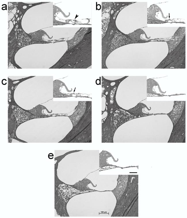
Photomicrographs of the UBT in: (a) normal hearing; and (b) 2 week; (c) 4 week; (d) 6 week; (e) and 10 week deafened rat cochleae. Note the absence of hair cells in the deafened cochleae, and the gradual degeneration of peripheral processes and SGNs. Scale bar = 100 μm. The inset in each micrograph illustrates the organ of Corti/basilar membrane at higher magnification. The arrowhead in the normal cochlea (a) illustrates the presence of outer hair cells. Arrows in the 2 and 4 week deafened cohorts (b & C) illustrate the structure of the organ of Corti in cochleae where the hair cells have undergone degeneration. In cochleae deafened for 6 and 10 weeks (d & e) the structure of the organ of Corti has completely degenerated. (Scale bar = 50 μm).