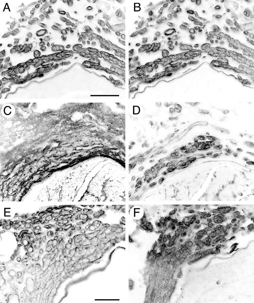
Figure 4.

Characterization of ERR basal laminae components. Transverse sections of 28 week regenerated sciatic nerve 3mm proximal to the coaptation were immunolabeled for (A) laminin and (B) perlecan. Colocalization of perlecan and laminin is consistent with that in basal laminae in normal nerve. Chondroitin-4-sulfate proteoglycan (CS4-PG) immunoreactivity was found surrounding the ERR minifascicles (C) while chondroitin-6-sulfate proteoglycan (CS6-PG) occupied the inner aspect of the minifascicles, more closely associated with the extraneural axons (D). NG2 immunolabeling circumscribed the ERR minifascicles, representing a subset of the CS4-PG distribution (E). Versican was restricted to the interior of the ERR sheaths and colocalized with CS6-PG immunolabeling (F). Scale bars: A-D, 50 μm; E-F, 25μm.
