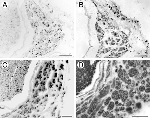
Figure 5.

Identification of myelinated motor and sensory axons within ERR minifascicles. Transverse sections of 28 week regenerated sciatic nerve 3mm proximal to the coaptation were immunolabeled for the specific sensory and motor axon markers, CGRP (A) and ChAT (B), respectively. ERR minifascicles contain both sensory and motor axons. The identification of S100 immunopositive Schwann Cells (C) and myelination with Sudan Black (D) indicates that many axons within the ERR minifascicles are well organized and ensheathed by differentiated Schwann cells. Scale bars: A-D, μm.
