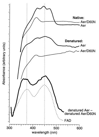
Figure 2.
Absorbance spectra of wild-type Aer and an FAD-binding mutant. His-tagged Aer proteins were solubilized in lauryl maltoside and were purified as detailed in Materials and Methods. Absorbance measurements were made with an Hitachi U-3300 UV/VIS spectrophotometer connected to a microcomputer. uv solutions software (Hitachi Instruments, San Jose, CA) was used to obtain the differential spectrum shown at the bottom of the figure. The vertical dashed lines mark the two local absorbance maxima of FAD.

