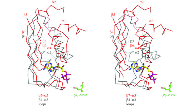
Figure 6.

Cα-trace overlay for part of the N-terminal domains of the LmMK and RnMK structures. The Cα trace and labels for LmMK are gray, and for RnMK red. The substrate, (R)-MVA is from the LmMK structure and shown as sticks colored green for C, red for O. The ATP (also in stick-mode, colored C yellow, N blue, O red, P purple) is from the RnMK structure. Selected elements of secondary structure are labeled and colored according to the structure, grey LmMK, red RnMK.
