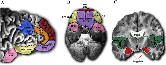
Figure 1. Regions Associated with Normal and Atypical Social Behaviour.
(A) Medial and lateral view of the PFC.
(B) View of the ventral surface of the PFC and temporal poles.
(C) Coronal slice illustrating the amygdalar and insular cortex.
See also Table 1.
ACC, anterior cingulate cortex; dlPFC, dorsolateral PFC; MFd, medial PFC; oMFC, orbitomedial PFC; TP, temporal pole; vlPFC, ventrolateral PFC; vmPFC, ventromedial PFC.

