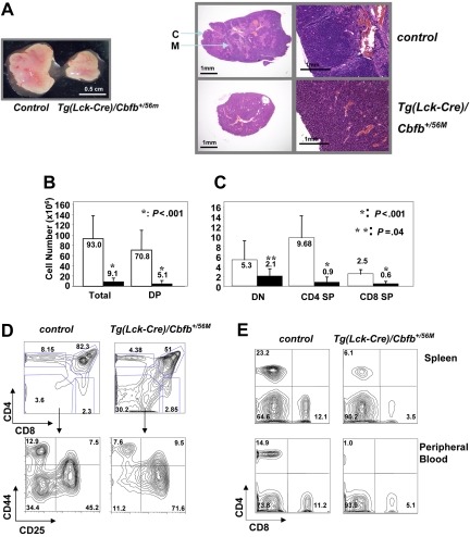
Figure 2.
T-cell developmental defects in the adult Tg(Lck-Cre)/Cbfb+/56M mice. (A) Tg(Lck-Cre)/Cbfb+/56M mice had smaller thymi (right) and their thymic architecture appeared to be homogeneous, as compared to the control mice (left). C indicates cortex; M: medulla. Left panel (gross view): pictures were taken with an Olympus SZ-40 (Tokyo, Japan) dissecting microscope camera with zoom setting at 2.5×. For the panel on the right, the 2 pictures on the left were taken with an Olympus SZ-40 dissecting microscope camera with zoom setting at 4×; the 2 pictures on the right were taken with a Nikon ECLIPSE E800 (Tokyo, Japan) microscope at 100× (10×/0.45 NA objective and 10× eyepiece). (B) Total and DP thymocyte numbers from adult thymi are graphed. (C) Numbers of DN, CD4+ SP, and CD8+ SP thymocytes are graphed. For panels B and C: □, littermate controls (n = 9); ■, Tg(Lck-Cre)/Cbfb+/56M mice (n = 10). Statistically significant P values (Student t test) are indicated. (D) Representative contour plots of cell surface marker expression of thymocytes from Tg(Lck-Cre)/Cbfb+/56M and littermate control mice. The cells were stained with CD4, CD8, CD44, and CD25. The top panels show CD4 and CD8 distribution, and the bottom panels show CD44 and CD25 staining of DN cells. (E) Representative contour plots of CD4 and CD8 expression of spleen and peripheral blood cells from Tg(Lck-Cre)/Cbfb+/56M and littermate control mice.

