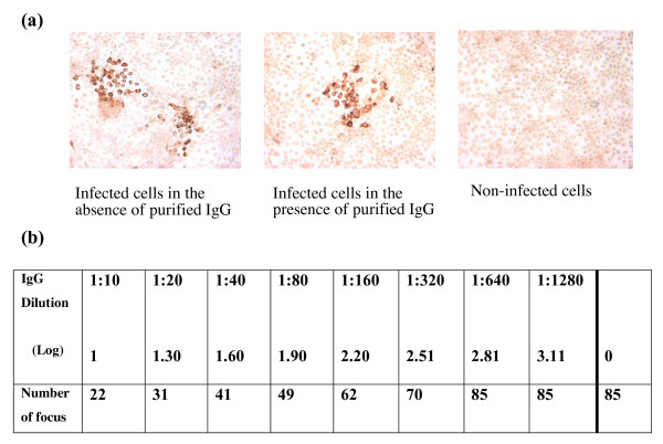
Figure 1.
(a) Typical pictures of HCVcc-infected Huh-7 cells observed under a light microscope (×40), showing the absence (on the right) and presence (on the left) of focus-forming units (FFU). (b) A sample-based neutralizing assay, showing the number of FFU in Huh-7 cells following serial dilutions of purified IgG from a HCV-positive serum sample. Neutralizing anti-HCV antibodies titers were expressed as the highest log dilution of IgG producing a 50% reduction in plaque count, as compared with controls in which the dose of virus was known.

