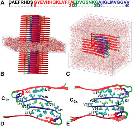
FIGURE 1.
MD simulations of Aβ9–40 protofilaments. (A) Aβ1–40 sequence and major structural elements: the unstructured N-terminal region (black), the N- (red) and C-terminal β-strands (blue), and the loop region (green). (B) Infinite-periodic fibril with solvent-filled simulation box. (C) Solvated 12-peptide fibril segment. (D,E) Top views of infinite fibrils with C2z (D) and C2x (E) topologies after 10 ns of MD.

