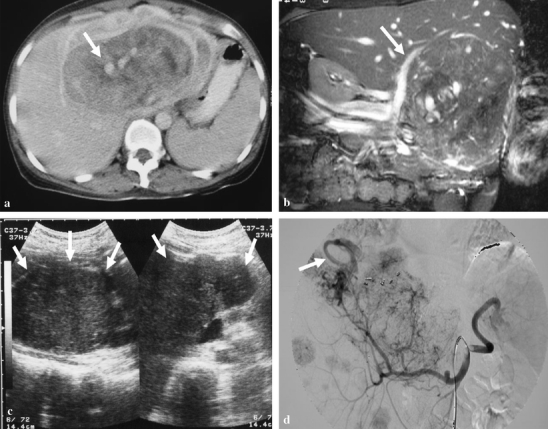
Figure 1.

A 37-year-old woman (case 3) presented with fever and palpable abdominal mass. (a) The axial view of contrast-enhanced CT scans on portal venous phase shows a huge hepatic tumor at the left hepatic lobe with heterogeneous enhancement. Notice the engorged vessels within the tumor are vividly identified (arrow). (b) The MR coronal Tru FISP, fast imaging with steady-state precession. (TR/TE/FA = 4.3/2.1/72°) shows engorged vessels in the tumor. The right portal vein (arrow) is displaced by the tumor. (c) After 6 months of extended left lobectomy, the abdominal ultrasonography reveals a huge recurrent tumor (arrows) in the previous location of left hepatic lobe, and numerous smaller tumors in the right lobe. (d) Celiac angiography also demonstrates the recurrent huge tumor and other multiple smaller ones in the right lobe of liver. Note the early drainage vein (arrow).
