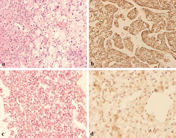
Figure 2.

Microscopic appearance of the hepatic angiomyolipoma in case 3. (a) The primary tumor is composed of polygonal to spindle cells arranged in solid sheets or trabecular pattern with endothelial lining. Some of the tumor cells have eosinophilic cytoplasm, and some have large fat vacuoles. Some of the nuclei are bizarre, and some have large eosinophilic nucleoli (H&E stain, original magnification ×100). (b) The tumor cells are strongly immunoreactive for HMB-45 (original magnification ×100). Recurrent tumor was noted 6 months later, and the patient received fine needle aspiration biopsy. (c) Microscopically, it shows tumor cells with clear to ample eosinophilic cytoplasm arranged in trabecular pattern (H&E stain, original magnification ×40). (d) Immunohistochemical staining shows the tumor cells are also positive for HMB-45 (original magnification ×200).
