
Figure 6.
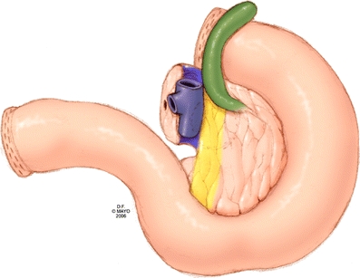
The specimen is carefully marked for all margins, including the venous segment margin. The portal vein groove and the retroperitoneal margin should be inked, and the venous segment should be evaluated histologically for malignant invasion (posterior view).