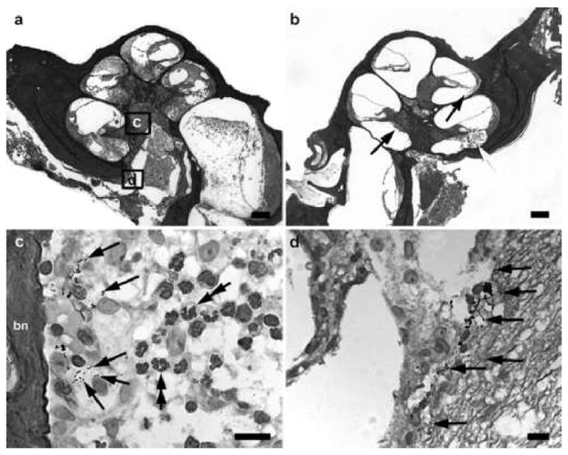
Figure 3.

Lower power H & E photomicrographs illustrating the implanted (a) and contralateral control (b) cochleae of a rat 48 hours following inner ear inoculation of 1 x 10³ CFU S. pneumoniae. This animal exhibited clinical and histological (CNS) evidence of meningitis. Extensive labyrinthitis of the inoculated left ear involved all three scalae. In contrast, the contralateral cochlea exhibited a less severe labyrinthitis with infection predominantly localized to the scala tympani. Higher power photomicrograph of Gram stain from the modiolus (c) and the internal acoustic meatus (d) implanted left cochlea illustrates the presence of bacteria (arrows). The approximate location of the higher power micrographs (c,d) are illustrated in (a) and (b). bn: bone. Scale bar: (a) & (b) 200 μm; (c) & (d) 10 μm.
