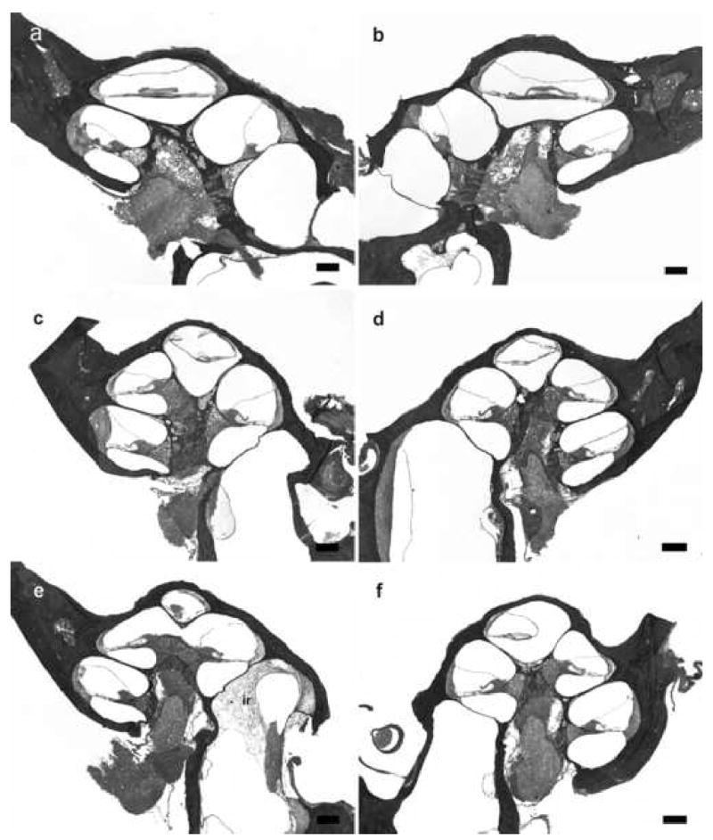
Figure 5.

Lower power H & E photomicrographs illustrating representative cochlear histology in acutely implanted non-meningitic animals. Left (a) and right (b) cochleae 120 hours after following IP inoculation of 4 x 10⁶ CFU S. pneumoniae. No inflammatory cells and bacteria were evident within both cochleae. Left (c) and right (d) cochleae 120 hours after middle ear inoculation of 3 x 10⁴ CFU S. pneumoniae. Similarly, no inflammatory cells and bacteria were evident within the cochleae. Left (e) and right (f) cochleae 120 hours following inner ear inoculation of 1 x 10³ CFU S. pneumoniae. Inflammatory cells response (ir) was evident in the basal turn of the ipsilateral cochlea. There were no inflammatory cells and bacteria in the other parts of cochlea or in the contralateral cochlea. There were no evidence of trauma to the OSL and the modiolus from acute insertion of the scala tympani electrode array. Reissiner’s membrane defects in some cochlear turns as seen in micrograph (e), were most likely to be artifacts from the histology preparation. An absence of organ of Corti was observed in the basal turn in the ipsilateral acutely implanted cochleae (a) and (e). The loss of organ of Corti did not alter the outcome of infection in these two animals. Scale bar: (a-f) 200 μm.
