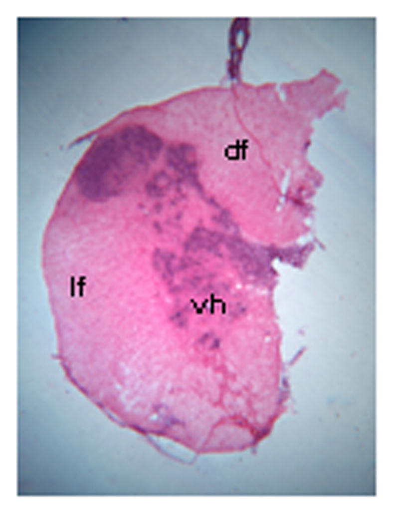
Figure 1.

Photomicrograph showing an example of the extent of a hemisection lesion in a hemisected rat, one week post injury; note that one half of the spinal cord is missing with the exception of some slight sparing of the dorsal funiculus. df = dorsal funiculus; lf = lateral funiculus; vh = ventral horn. Magnification: 25X.
