
Figure 3.
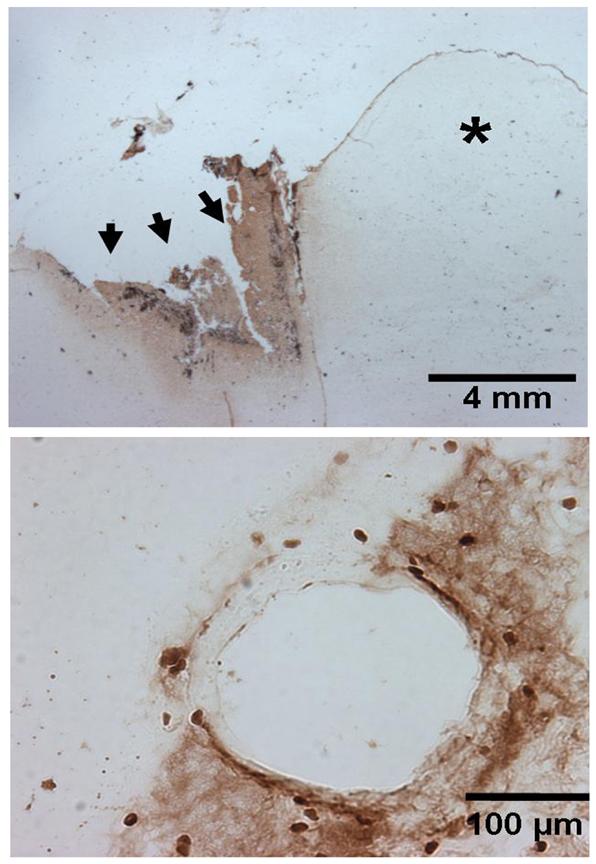
The representative figure (n=4) in the upper panel shows IgG staining in a horizontal section of the brain 24 hours after the frontal lobe surgical injury. There is increased IgG staining surrounding the surgically-induced brain injury (marked by arrows) on the ipsilateral side as compared to the unaffected contralateral side indicated by the asterisk (*). The lower panel shows an individual affected blood vessel at high magnification depicting disruption of blood brain barrier as indicated by IgG staining. Scale represents 4 mm and 100 μm in upper and lower panels respectively.