
Fig. 6.
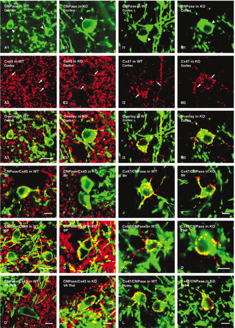
Laser scanning confocal immunofluorescence of Cx43 and Cx47 association with CNPase-labeled oligodendrocytes in various brain regions of wild-type (WT) and Cx32 knockout (KO) mice. A: Confocal double labeling in cerebral cortex of WT mouse showing two CNPase-positive oligodendrocytes (A1, arrowheads) with Cx43-positive puncta (A2, arrows) on the cell bodies (yellow in overlay image, A3). B–D: Confocal micrographs from WT mouse showing CNPase (green) co-association with Cx43 (red) as seen by yellow in overlays from striatum (B), globus pallidus (C), and ventroanterior thalamic nucleus (D). E: Confocal double labeling in cerebral cortex of Cx32 KO mouse showing CNPase-positive oligodendrocyte (E1, arrowheads) with Cx43-positive puncta (E2, arrows) on its surface (yellow in overlay, E3). F–H: Confocal micrographs from Cx32 KO mouse showing CNPase (green) co-association with Cx43 (red), as seen by yellow in overlays from striatum (F), globus pallidus (G), and ventroanterior thalamic nucleus (H). I: Confocal double labeling in cerebral cortex of WT mouse showing a CNPase-positive oligodendrocyte (I1, arrowhead) with Cx47-positive puncta (I2, arrows) on the cell body (yellow in overlay, I3). J–L: Confocal micrographs from WT mouse showing CNPase (green) co-association with Cx47 (red), as seen by yellow in overlays from striatum (J), thalamus (K), and hypothalamus (L). M: Confocal double labeling in cerebral cortex of Cx32 KO mouse showing a CNPase-positive oligodendrocyte (M1, arrowhead) with Cx47-positive puncta (M2, arrows) on its surface (yellow in overlay, E3). N–P: Confocal micrographs from Cx32 KO mouse showing CNPase (green) co-localization with Cx47 (red), as seen by yellow in overlays from striatum (N), thalamus (O) and hypothalamus (P). Scale bars = 10 μm in A–C; 5 μm; in D–H,I–N,O,P.