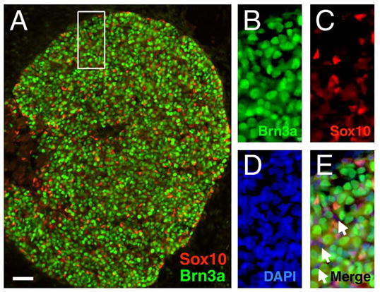
Figure 1. Cellular expression of Brn3a in the embryonic trigeminal ganglion. Guinea-pig anti-Sox10 (Maka et al., 2005) and rabbit anti-Brn3a were used to perform immunofluorescence as previously described (Fedtsova and Turner, 1995).

(A) In the E13.5 trigeminal, the majority of cells are Brn3a-expressing neurons. The relatively small population of differentiating glia present at this stage are identified by small nuclei and the expression of Sox10.
(B–E) An enlarged view shows mutually exclusive cellular expression of Brn3a and Sox10 (boxed area in A). Occasional cells express neither antigen (arrows, E), and may represent nucleated blood cells.
