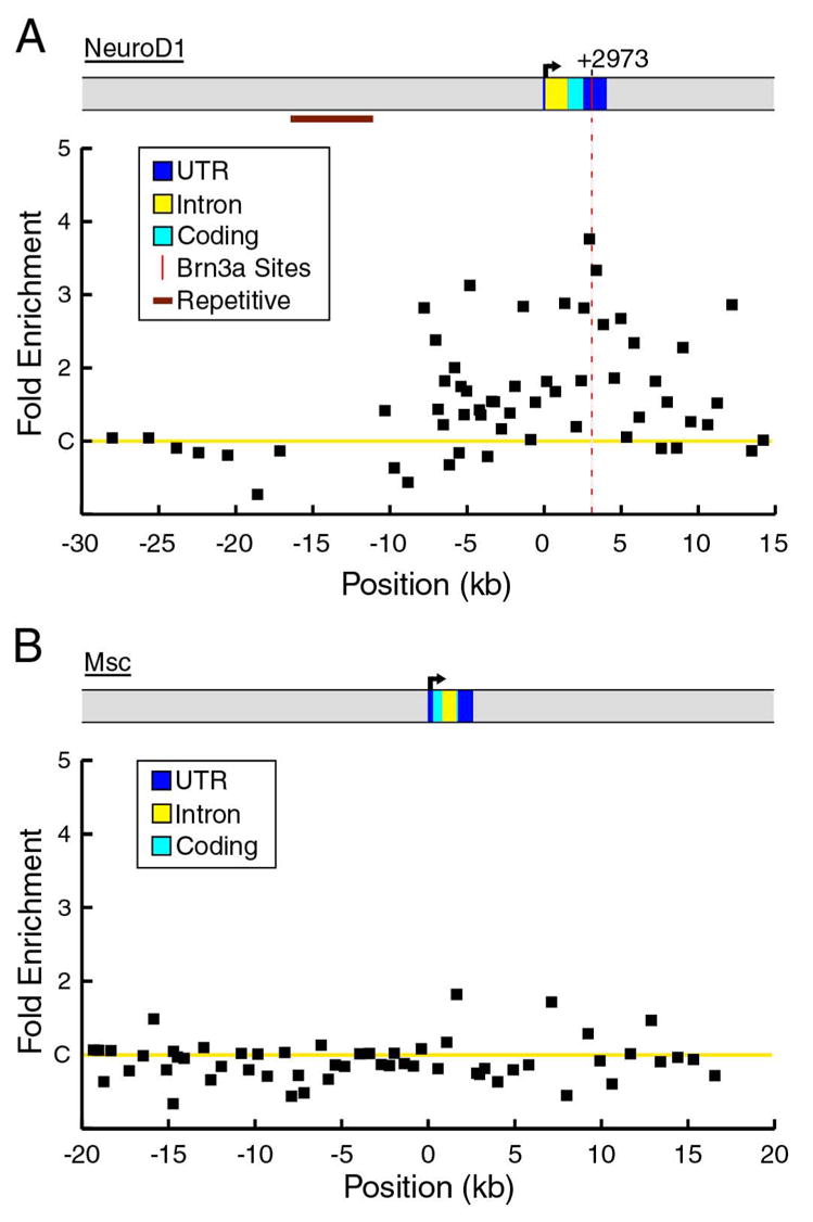
Figure 3. ChIP analysis of Brn3a binding to the NeuroD1 and Msc loci in E13.5 trigeminal ganglia.

(A) In vivo binding of Brn3a to the NeuroD1 locus. A peak of enrichment in a region of the 3′ UTR conserved between the mouse and human genomes includes a Brn3a consensus site at +2973 (difference from controls, p=0.008). The region extending from −16 kb to −11 kb consists of repetitive sequence and was not included in the analysis. ChIP primer pairs used for the NeuroD1 locus appear in Table S4.
(B) Brn3a ChIP analysis of the Msc locus from −20kb to +20 kb relative to the start of transcription reveals no significant in vivo binding of Brn3a. Average enrichment values from three independent ChIP assays are shown for both loci. Primer pairs used in ChIP assays of the Msc locus appear in Table S5.
