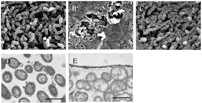
Figure 4.
SEM and TEM analysis of V. fischeri cells expressing rscS1. The production of an extracellular matrix was examined by SEM from (A) vector-control wild-type cells (KV1844), (B) rscS1-expressing wild-type cells (KV1956) and (C) rscS1-expressing sypN mutant cells (KV1992) and by TEM analysis of (D) vector control and (E) rscS1-containing wild type cells stained with ruthenium red. Bars represent 1 μm.

