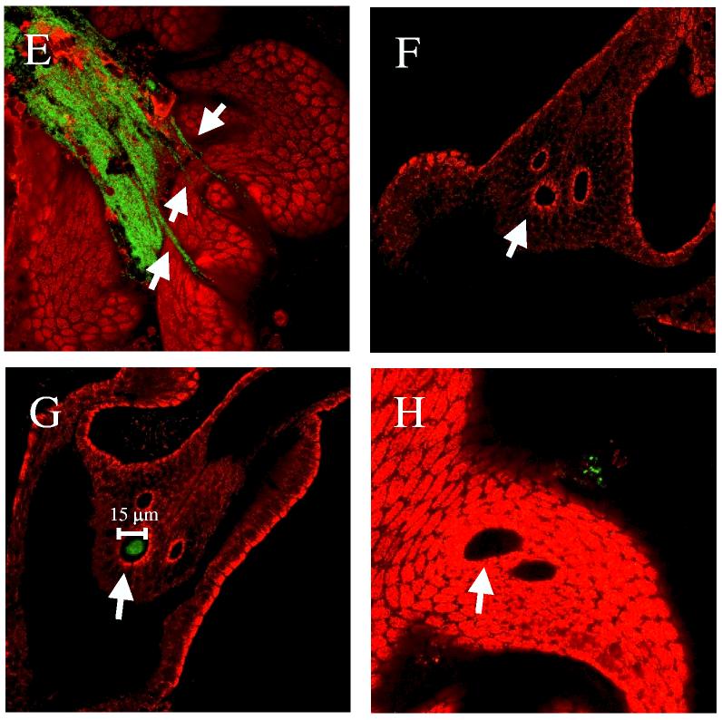
Figure 7.
Aggregate formation on the light organ by V. fischeri. Newly hatched juvenile squid were inoculated with GFP-labeled bacteria. After 2 to 6 h, animals were stained with Cell Tracker Orange (red color) and the light organs examined by confocal microscopy. Representative images of aggregated V. fischeri cells at or near a light organ pore are shown. Animals were inoculated with (A) ES114 (KV1066), (B) rscS mutant cells (KV1339), (C) wild-type cells carrying the vector control (KV2685), (D and E) wild-type cells carrying rscS1 (KV2688), (F and G) rscS1-expressing sypN mutant cells (KV2689), and (H) sypN mutant cells carrying vector (KV2686). Arrows indicate pores of the light organ.


