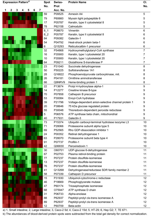
Figure 4.

Clustering of identified protein spots according to the expression levels in analyzed samples (continuation of Figure 3). Identified protein spots grouped in different clusters (cluster 5–12) according to the expression level in different samples. Triplicate spot intensities are shown by a colour range; bright red, black, and bright green represent high, average, and low levels of protein expression.
