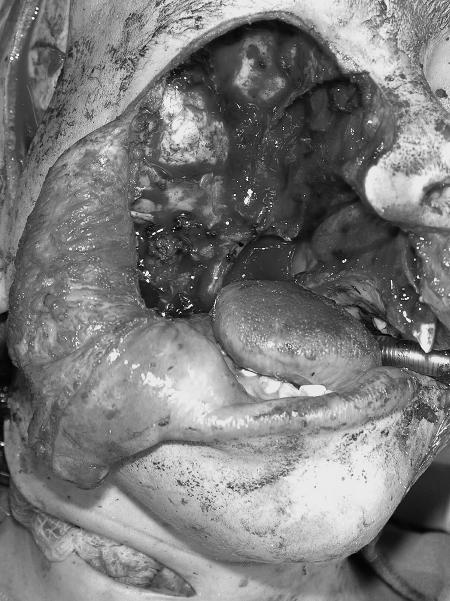
Figure 1.
Intraoperative photograph following resection of parameningeal recurrent rhabdomyosarcoma. Note the exposed temporal dura in continuity with the oropharynx. (Figure is property of The Department of Neurosurgery, The University of Texas M.D. Anderson Cancer Center and is used with permission.)

