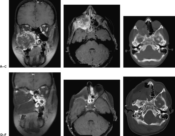
Figure 5.
Preoperative (A) coronal and (B) axial contrast-enhanced MRI as well as preoperative (C) CT reveal a large recurrent rhabdomyosarcoma centered in the maxillary sinus with extension into the orbit, sphenoid wing, and infratemporal fossa. Postoperative (D) coronal and (E) axial contrast-enhanced MRI as well as postoperative (F) CT show the extent of soft-tissue and skull base resection and the subsequent reconstruction using the VRAM flap. (Figure is property of The Department of Neurosurgery, The University of Texas M.D. Anderson Cancer Center and is used with permission.)

