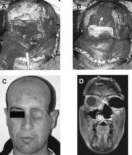
Figure 2.
Orbital reconstruction following subcranial resection in a 28-year-old patient with T4AN0M0 squamous cell carcinoma. (A) The orbita was reconstructed with temporalis muscle rotational flap. The skull base was reconstructed with a double-layer fascia lata. (B) The periorbit was reconstructed with the nasofronto-orbital bone segment and with titanium mesh. Wrapping of the frontonaso-orbital segment was accomplished with a pericranial flap (arrow). (C) A picture of the patient 12 months after surgery. (D) A postoperative T1 MRI shows the area of reconstruction.

