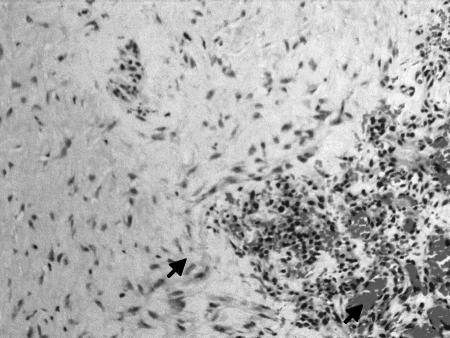
Figure 5.
Neovascularization of the fascia lata graft 12 months after surgery in a case with recurrent tumor (H & E staining). Microscopical examination shows fibroblasts embedded in a dense collagenous stroma (higher arrow). Note the presence of neovascularized channels lined by endothelial cells (lower arrow). The graft has been replaced with a viable dense fibrocollagenous tissue. (20 × magnification.)

