
Figure 5.
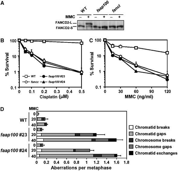
Chicken DT40 cells in which the FAAP100 gene was inactivated display hallmark features of FA cells. (A) Immunoblotting shows that DT40 cells nullizygous for FAAP100 have no detectable monoubiquitinated FANCD2. The whole-cell lysates prepared from wild-type (WT) and FANCL-deficient (fancl) cells were included as positive and negative controls, respectively. Cells were either treated with MMC (500 ng/ml) for 6 h or left untreated. (B, C) The colony survival assays show that faap100-null cells are hypersensitive to DNA crosslinking drugs cisplatin and MMC. The percentage of the surviving colonies following treatment with the drugs at the indicated concentrations are shown. Two clones of faap100 cells were included in the analysis. The data shown are mean±standard deviation of at least three separate experiments. Wild-type and FANCC-knockout (fancc) cells were used as controls. (D) The chromosomal breakage analysis revealed that faap100-null cells have increased chromosomal abberations. Wild-type and two clones of faap100 cells were treated with MMC (20 or 40 ng/ml) for 24 h or left untreated. Chromosome aberrations were counted blindly using coded slides as described (Yamamoto et al, 2003). The counting was performed only on large chromosomes (total 12). Error bars represent standard error of total aberrations per metaphase. At least 150 (for wild-type) or 50 cells (for faap100) were scored for each preparation.