
Figure 1.
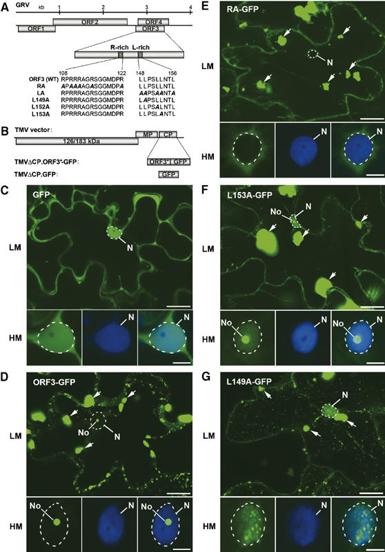
ORF3 protein domains involved in nuclear localization and cRNP formation. (A) Schematic representation of the GRV genome with protein sequences of the wild-type (WT) and mutant ORF3 R- and L-rich domains. (B) Representation of the TMV-based vector for expression of WT and mutated ORF3 (ORF3*)-GFP fusion proteins or GFP alone replacing the TMV CP gene. (C–G) Intracellular localization of free GFP (C), ORF3-GFP (D) and the mutant GFP fusions: RA-GFP (E), L153A-GFP (F) and L149A-GFP (G) expressed from a TMVΔCP vector in epidermal cells and determined by CLSM. Each set of GFP images presents a whole cell at low magnification (LM) and a nucleus at high magnification (HM) showing GFP image (left-hand panel), DAPI staining (centre panel) and overlaid GFP and DAPI images (right-hand panel). Free GFP localizes to the nucleus (largely excluded from the nucleolus) and cytoplasm (C) whereas ORF3-GFP localizes to the nucleolus and cytoplasmic inclusions (indicated by arrows) (D). RA-GFP is largely excluded from the nucleus (E), L153A-GFP is nuclear with strong localization to the nucleolus (F) and L149A-GFP is nuclear with strong accumulation in small nuclear bodies (G). No, nucleolus; N, nucleus (shown by dashed line). Scale bars, 20 μm (LM) and 5 μm (HM).