Figure 4.

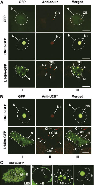
Effect of ORF3 protein on the integrity and localization of CBs. (A, B) Confocal images of epidermal cells infected with TMVΔCP expressing ORF3-GFP, L149A-GFP and free GFP (as a control) and immunostained using antibodies to coilin (A) and U2B″ (B). In ORF3-GFP-infected cells, coilin and U2B″ localize with the ORF3 protein to nucleoli, whereas in L149A-GFP-infected cells, these proteins are found in multiple small bodies (CBLs). CBs are detected in GFP-expressing cells but not in ORF3-GFP cells. Panels: I—GFP images; II—fluorescent (red) antibody labelling; III—overlay images. CB, Cajal bodies; CBL, CB-like structures (shown by arrowheads); No, nucleoli; N, nuclei (indicated by dashed lines according to DAPI staining). Chl, chloroplasts showing natural red autofluorescence. Scale bars, 5 μm. (C) Intranuclear localization of the ORF3-GFP protein in a pseudo-time course of ORF3-GFP infection. The left panel (LES, for lesion) shows a GFP image of a whole virus-infected lesion with cells corresponding to different stages of infection. Cells at the front of the infection site represent early infection events (E), whereas those cells in the centre of the infection site represent late events (L). ‘Early' event cells show fluorescence mainly in the perinuclear region; intermediate (middle) stage cells (M) show accumulation of fluorescence in multiple subnuclear bodies, similar to the CBLs that merge with the nucleolus at later stage (L). Scale bars, 5 μm, except for lesion in (C) (left panel)—200 μm.
