
Figure 5.
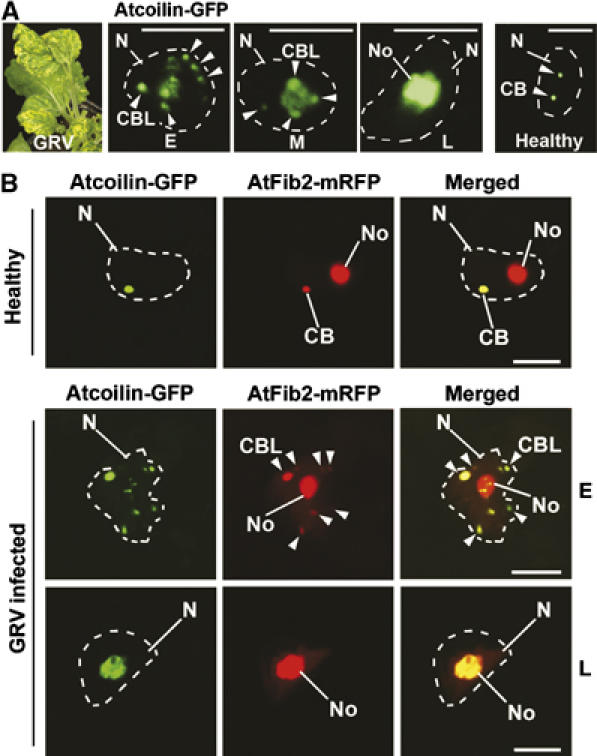
Fusion of CBLs with the nucleolus in GRV-infected plants. (A) Localization of Atcoilin-GFP delivered by Agrobacterium in a pseudo-time course of GRV-YB infection from early (E), middle (M) and late (L) stages of the infection compared to a healthy cell. GRV-YB infection induces yellow blotch symptoms (left panel) at the front of systemic infection and therefore acts as a visible marker of infection progression. Atcoilin-GFP fluorescence in cells at different stages of GRV infection shows multiple CBLs merging with the nucleolus, whereas in healthy cells, CBs remains distinct from the nucleolus (right panel). (B) Subnuclear colocalization of Atcoilin-GFP and AtFib2-mRFP delivered by Agrobacterium into healthy or GRV-YB—infected plant cells at early (E) and late (L) stages of GRV infection. In healthy plant cells, AtFib2 labels both the CBs (usually 1–3 per nucleus) and the nucleolus, whereas Atcoilin colocalizes with AtFib2 only in CBs. At early stages of GRV infection (E), AtFib2 colocalizes with Atcoilin in multiple CBLs, which merge with the nucleolus at late stage (L). CB, Cajal bodies; CBL, CB-like structures (shown by arrowheads); No, nucleoli; N, nuclei (shown by dashed lines according to DAPI staining). Scale bars, 5 μm.