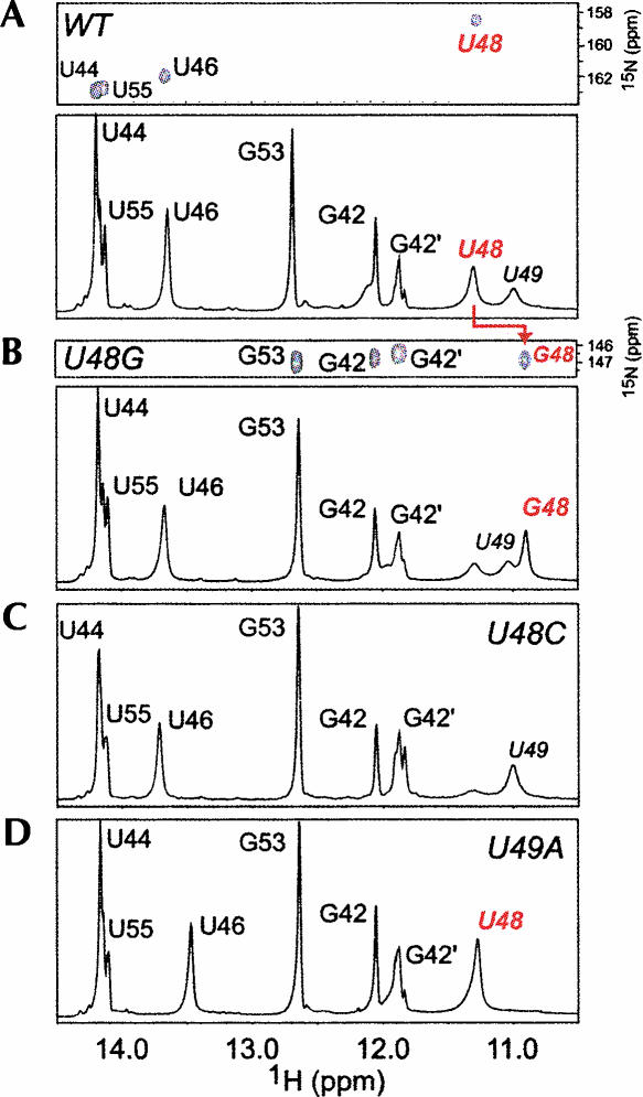
FIGURE 5.
Imino proton regions of 1D jump–return echo spectra acquired at 10°C, pH 6.0 for SL2 variants, with 1H-15N-HSQC spectra shown for 13C,15N-[U]-labeled WT SL2 and 13C,15N-[G]-labeled U48G SL2, as well. (A) WT SL2 (see Fig. 3B); (B) U48G SL2; (C) U48C SL2; and (D) U49A SL2. G42 and G42′ represent alternative conformations for the terminal G42-C56 base pair. The immediately adjacent U55 resonance is also doubled, indicative of heterogeneity at the base of SL2.

