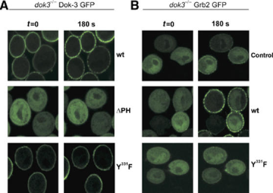
Figure 5.

Dok-3 is permanently localized at the plasma membrane and is essential for stimulation-dependent recruitment of Grb2. (A) Dok-3-deficient DT40 mutants were transfected with expression constructs encoding fusion proteins between the green fluorescence protein (GFP) at the C terminus and either wild-type Dok-3 (upper row), Dok-3ΔPH (middle row) or Dok-3 Y331F (lower row) at the N terminus. Subcellular localization of Dok-3/GFP fusion proteins in resting (t=0) or BCR-activated (180 s) cells (left and right images) was visualized by confocal laser scanning microscopy. (B) Dok-3−/− DT40 cells expressing a Grb2/GFP fusion protein were transfected with either empty control vector (upper row) or expression vectors encoding wild-type or Y331F Dok-3 mutants (middle and lower rows). Subcellular Grb2 localization was analyzed as in (A). The Ca2+ signaling function of GFP fusion proteins was tested separately (data not shown).
