
Figure 5.
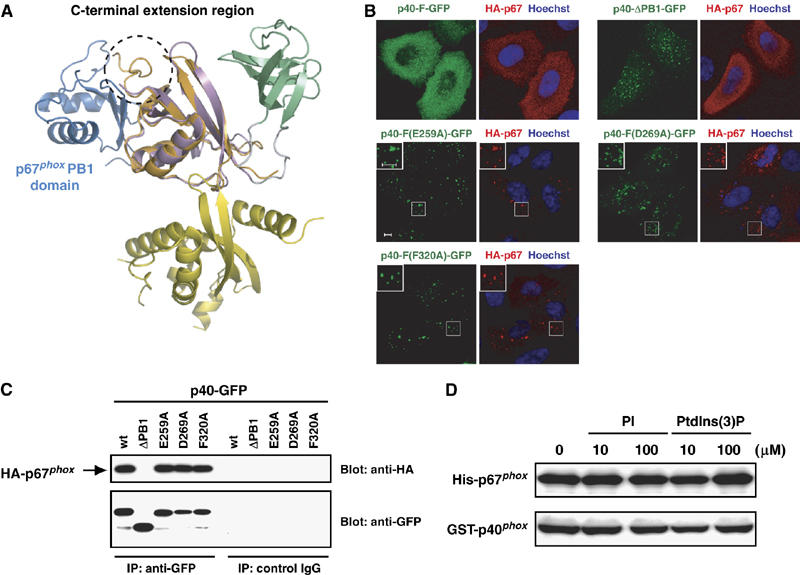
Dual roles for the PB1 domain of p40phox. (A) Ribbon diagram of the superposition of p40phox/p67phox PB1 heterodimer (blue is p67phox PB1 and orange is p40phox PB1; PDB code 1OEY) onto that of the full-length p40phox (color coded as in Figure 1A). A C-terminal extension region is shown in dashed circle. (B) Subcellular distribution of GFP-tagged p40phox and HA-tagged p67phox in transiently transfected HeLa cells. Left panel, distribution of GFP-tagged p40phox in fixed HeLa cells (green); right panel, distribution of endogenous HA-tagged p67phox (red) and Hoechst staining of the nucleus (blue) in the same field of fixed HeLa cells. The insets show the magnified views. Scale bar, 5 μm. (C) Proteins of lysates prepared from HeLa cells expressing both p40phox and p67phox were immunoprecipitated (IP) with the anti-GFP or control IgG, and then analyzed by immunoblot (Blot) with the anti-HA or anti-GFP antibodies. (D) In vitro interaction between purified GST-p40phox and His-tagged p67phox in the presence or absence of PtdIns(3)P.