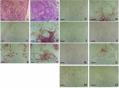
Fig 5.
Immunohistochemical evaluation of the lymphoid cells in the pre- and postvaccine (180 days) biopsies taken from a patient with inflammatory breast carcinoma (Table 2, patient 3; Fig. 4). (A) and (B) Hematoxylin and eosin staining to show the tumor cells infiltrating the skin. (C) and (D) T cells evaluated by CD43. (E) and (F) T cells evaluated by CD45Ro. (G) and (H) NK cells evaluated by CD57. (I) and (J) Macrophages evaluated by CD68. (K) and (L) B cells evaluated by CD20. (M) and (O) Leukocytes evaluated by CD15. Bar = 60 μm (A–F); bar = 100 μm (G–O)

