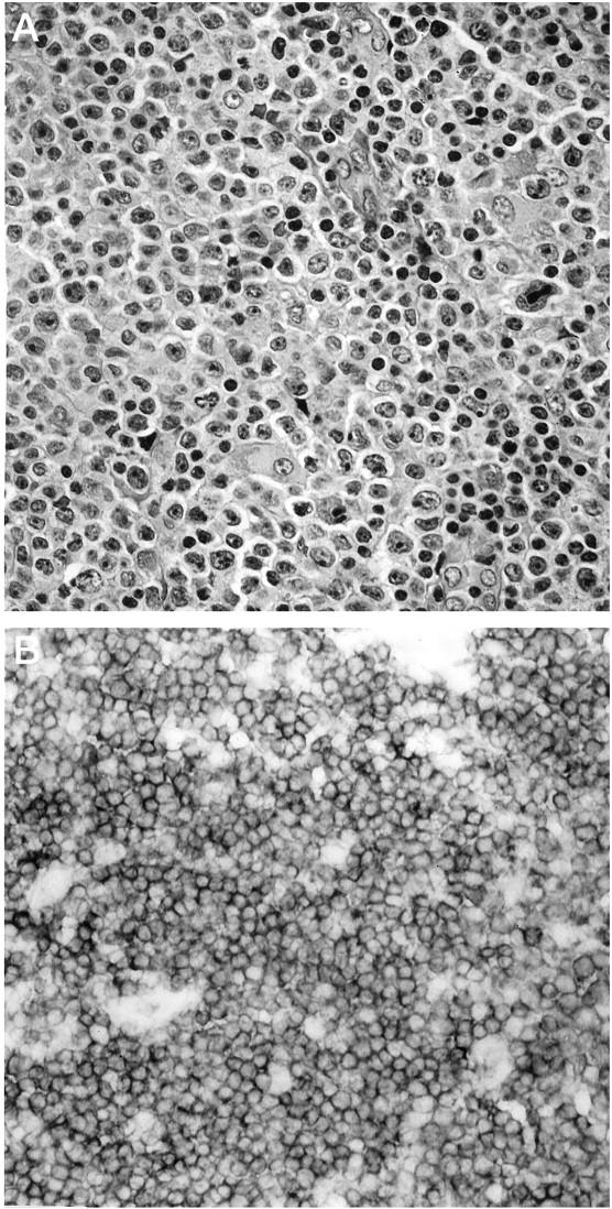
Figure 1.

Immunoreactivity for CD100 in a case of diffuse, large-cell non-Hodgkin’s lymphoma, T-cell type. A: Representative neoplastic tissue fixed in 10% buffered formaldehyde, paraffin embedded, sectioned, and stained with H&E. B: CD100 immunoperoxidase staining of frozen neoplastic tissue. Large neoplastic cells exhibit strong, uniform surface staining for CD100.
