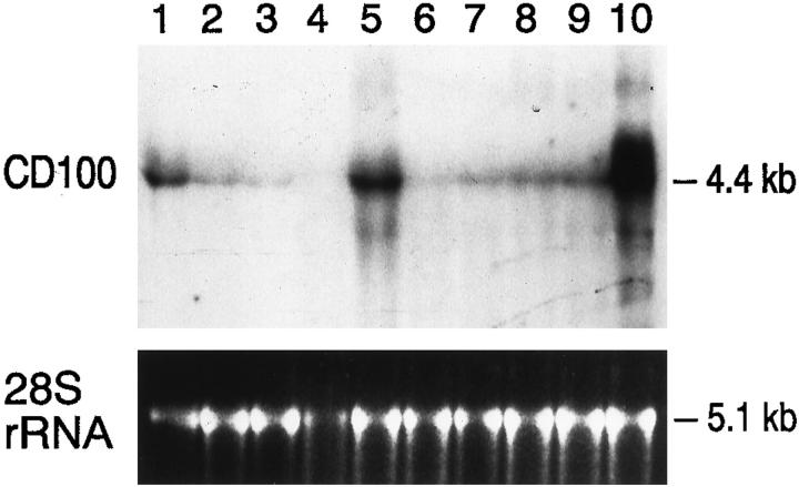
Figure 5.
CD100 mRNA expression in non-Hodgkin’s lymphomas. Northern blot containing RNA prepared from 1) activated T cells; 2) Rex (acute T-cell lymphoblastic leukemia cell line); 3) Raji (human Burkitt lymphoma cell line); 4) mantle cell non-Hodgkin’s lymphoma; 5) follicular lymphoma, predominantly large-cell type; 6) follicular lymphoma, predominantly small-cell type; 7) high-grade, small non-cleaved (Burkitt-like) lymphoma; 8) chronic lymphocytic leukemia (CLL)/small lymphocytic lymphoma (SLL); 9) reactive lymph node; and 10) T-cell non-Hodgkin’s lymphoma, diffuse, large-cell type. Each lane contains 20 μg of total RNA and was probed with the 4.3-kb CD100 cDNA insert. The position of the 4.4-kb CD100 mRNA is indicated. The bottom panel shows the ethidium-bromide-stained 5.1-kb 28 S rRNA corresponding to each of the above lanes. The neoplasms studied in lanes 5, 6, 7, and 10 correspond to those shown in Figures 3, 2, 4, and 1 ▶ ▶ ▶ , respectively.

