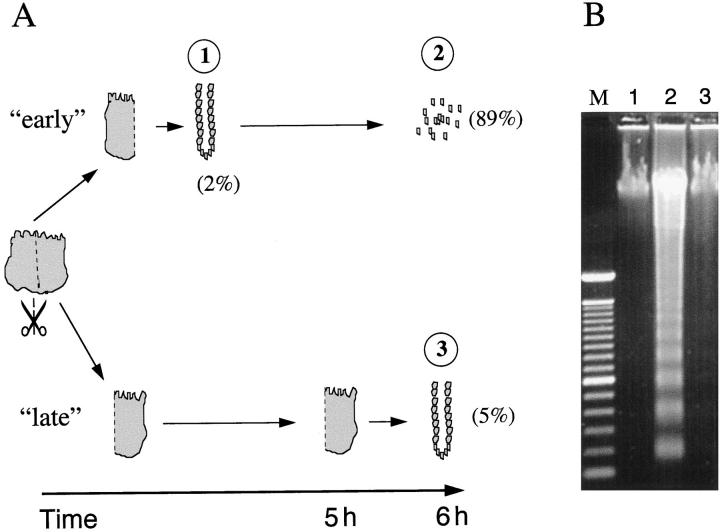
Figure 2.
ECM anchorage protects IECs from anoikis. A: Surgical specimens were split in half. IECs from one half were isolated immediately (method D) and subsequently incubated at 37°C (“early”). Tissue from the other half was incubated intact at 37°C, allowing IECs to maintain anchorage. After 5 hours, IECs from the second half (“late”) were isolated using the same protocol. The apoptotic index is indicated by the numbers in parentheses (%). The circled numbers correspond to the lanes of the gel in B. B: DNA, extracted after immediate processing (“early”) of the tissue (lane 1, IECs immediately after isolation; lane 2, IECs 6 hours after isolation) and after delayed processing (“late”) of the tissue (lane 3), was analyzed by electrophoresis (lane M, molecular mass marker). Data are representative of three experiments.

