
Figure 3.
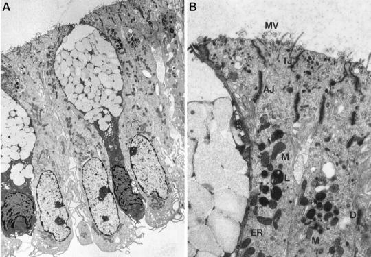
Lack of apoptosis in IECs isolated according to method D as determined by electron microscopy. A: Longitudinal cross section of purified crypt epithelial cells examined by transmission electron microscopy (magnification, ×6000). Note the fully preserved columnar shape of tightly joined colonocytes, the goblet shape of the mucus-containing cells, and the preserved basolateral location of the nuclei. B: Ultrastructure (magnification, ×48,000) of IECs, showing full preservation of cell-cell junctions, including tight junctions (TJ), adherent junctions (AJ), desmosomes (D), microvilli (MV), and organelles such as endoplasmic reticulum (ER), lysosomes (L), and mitochondria (M).