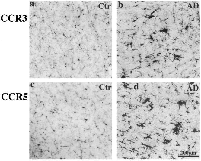
Figure 1.
CCR3 (7B11) and CCR5 (3A9) immunoreactivity in the inferior temporal lobes of a 58-year-old control patient and an 81-year-old AD patient with duration of illness for 12 years (PMIs less than 8 hours). CCR3 (a and b) and CCR5 (c and d) immunoreactivities are clearly seen on microglia of both cases. In the control case, the majority of cells stained are resting microglia, whereas in the AD brain both resting and reactive microglia cells are clearly stained, and some reactive microglia appear in clusters. All images have the same scale of magnification. Scale bar, 200 μm.

