
Figure 3.
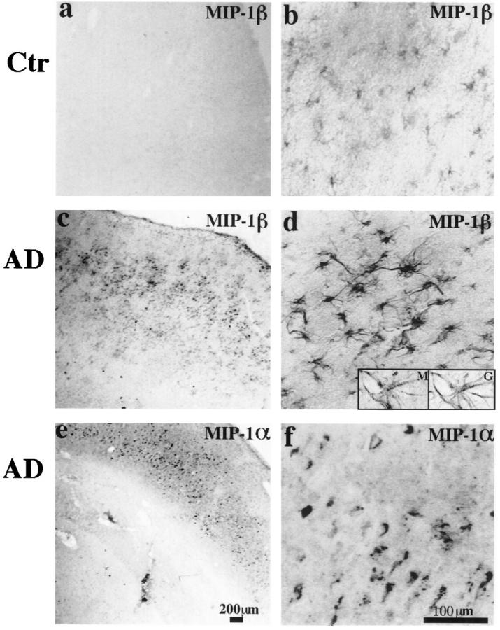
a to d: MIP-1β (1F12) immunoreactivity in the inferior temporal lobes of a 63-year-old control patient and a 58-year-old AD patient (PMIs less than 8 hours). In the control case, weak MIP-1β staining can be found on a small population of resting and some occasional reactive astrocytes. However, in the AD case, much more widespread and stronger astrocyte staining is observed, and most of the astrocytes appear to be reactive. Insets are high-power images (inverted images) of double staining of MIP-1β (M, Cy3) versus GFAP (G, bodipy), showing a MIP-1β-positive cell is also clearly positive for GFAP. e to f: MIP-1α (11A3) immunoreactivity in the hippocampal formation of an 84-year-old AD patient (PMI, 7 hours). A diffused pattern of neuronal staining can be seen. Both neurons and neuropil were stained. Some neurons showed increased expression of MIP-1α. This pattern of immunoreactivity can be completely blocked by preabsorption with MIP-1α protein. a, c, and e: Low-power images (scale bar, 200 μm); b, d, and f are higher-power images (scale bar, 100 μm).