
Figure 1.
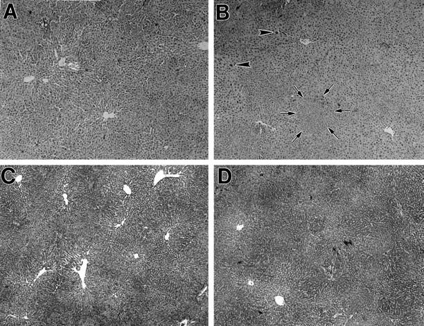
Liver histology of rats treated with retrorsine or retrorsine plus hepatocyte transplantation and partial hepatectomy. A: DPPIV− F344 rat 1 month after treatment with two doses of retrorsine. Note liver sinusoidal dilatation and moderate disorganization of hepatic lobular structure, but no evidence of acute or chronic inflammation (H&E stain). B: DPPIV− F344 rat treated with two doses of retrorsine as in A, followed by transplantation of 2 × 10 6 hepatocytes and two-thirds partial hepatectomy. The animal was sacrificed 2 months after cell transplantation, and histological analysis was performed. Note scattered foci of proliferating hepatocytes (one focus circumscribed by arrows). There is also marked variation in the size of hepatocytes, including some with very large nuclei (arrowheads). The lobular organization is improved compared to pretransplantation status, but this varies from animal to animal (H&E stain). C: Same as in A, using trichrome staining. Note moderate disruption of hepatic lobular structure, but no evidence of hepatic fibrosis. D: Same as in B, using trichrome stain. Note areas of focal proliferation of hepatocytes, but no evidence of margination between transplanted and endogenous hepatocytes, as well as absence of fibrotic changes. Original magnification: A through D, ×50.