
Figure 2.
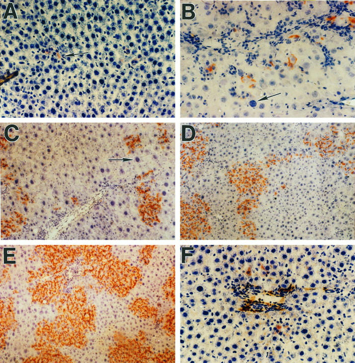
Proliferation of DPPIV+ hepatocytes in the liver of retrorsine-treated female DPPIV− mutant rats after partial hepatectomy. A: One day after transplantation of 2 × 10 6 hepatocytes. Scattered, isolated DPPIV+ cells are located in hepatic sinusoids near the portal triads (arrow). B: Four days posttransplantation. Transplanted cells are present in small clusters of two to four cells, suggesting several rounds of cell division. The arrow points to a megalocytic DPPIV− hepatocyte with a large hyperchromatic nucleus. C: Thirteen days posttransplantation. The size of DPPIV+ cell clusters is increasing, and clusters are now visible at a distance farther from the portal triads (mid-parenchyma). The arrow points to one of many megalocytic DPPIV− hepatocytes that become prominent after partial hepatectomy. D: One month posttransplantation. The average number of DPPIV+ cells in clusters continues to increase, and larger clusters are beginning to become confluent. Although there is variation from section to section and within sections, approximately 15 to 25% of total liver mass is now replaced by DPPIV+ cells. E: Two months posttransplantation. The size of DPPIV+ clusters continues to increase, and transplanted hepatocytes now constitute 40 to 60% of total hepatic mass. F: One month posttransplantation in the absence of partial hepatectomy. Under these conditions, there is very little proliferation of transplanted cells, which are visible as single cells or doublets primarily in the periportal region. Original magnifications: A, B, C, and F, ×200; D and E, ×100.