
Figure 5.
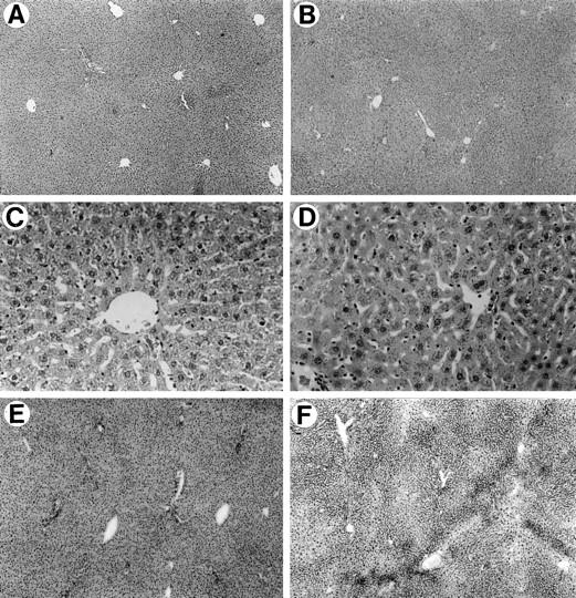
Histological and histochemical analysis of the liver 9 months after hepatocyte transplantation. A, C, and E: untreated DPPIV− rat liver. B, D, and F: retrorsine-treated DPPIV− rat liver 9 months after partial hepatectomy and transplantation of 1 × 10 6 DPPIV+ hepatocytes. A and B: H&E staining of formalin-fixed and paraffin-embedded tissue at ×50 magnification. C and D: H&E staining of formalin-fixed and paraffin-embedded tissue at ×200 magnification. E and F: DPPIV histochemical staining at ×50 magnification. Except for mild reduplication of bile ducts in B and D, the lobular structure and organization of the liver in the transplanted animal is essentially normal. E: Liver removed from this rat at the time of partial hepatectomy after retrorsine treatment but before cell transplantation. F: Liver from the same rat 9 months after transplantation of DPPIV+ hepatocytes.