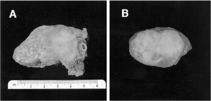
Figure 1.
Gross photographs of two VHL-associated pancreatic NETs. A: A 3-cm solid, well-circumscribed, tan-gray tumor (tumor 1) with prominent collagen stroma removed from patient 1 by partial pancreatectomy. B: A 1-cm NET (tumor 29) removed from patient 14 by enucleation. The tumor displays a prominent yellow color secondary to abundant lipid content.

