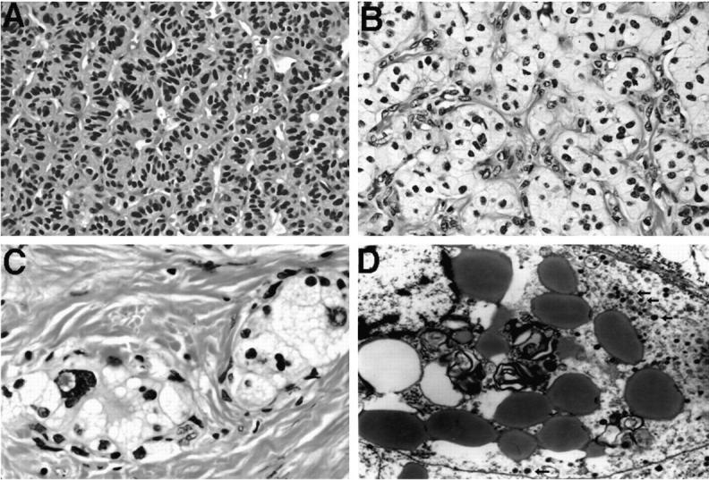
Figure 2.

Histology and electron microscopy of representative VHL-associated pancreatic NETs. A: Tumor 19 (patient 9) shows trabecular architecture and small NET cells with eosinophilic cytoplasm (H&E, ×400). B: Tumor 18 (patient 8) shows solid architecture, small vessels, and cells with prominent clear cytoplasm (H&E, ×400). C: Tumor 14 (patient 6) demonstrates nests of tumor cells with clear cytoplasm and focal nuclear atypia surrounded by stromal collagen bands (H&E, ×630). D: Electron micrograph of a “clear” cell from tumor 18 (patient 8); prominent lipid globules and myelin figures and small dense core granules (arrows). Magnification, ×8900.
