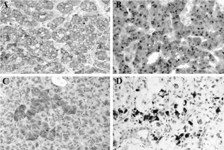
Figure 3.
Immunohistochemistry results in representative VHL NETs. A: Positive synaptophysin stain in tumor 19 (patient 9) (×400). B: Positive S100 stain in tumor 19 (patient 9) (×400). C: Focally positive pancreatic polypeptide stain in tumor 11 (patient 5) (×400). D: Focally positive insulin stain in tumor 14 (patient 6) (×400).

