Figure 4.
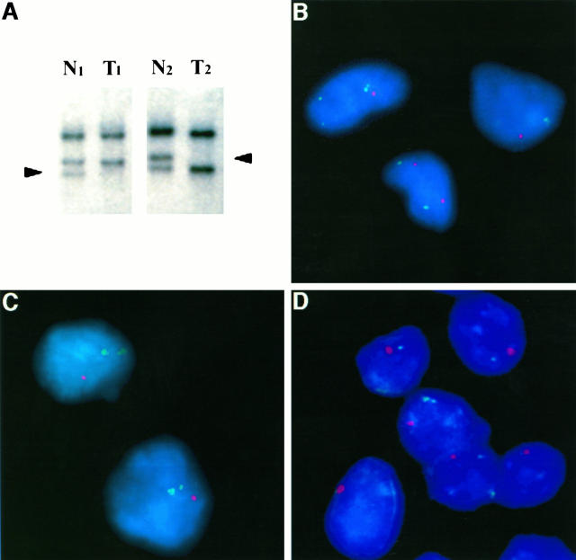
A: Detection of VHL gene (3p25.5) LOH by PCR-SSCP analysis in tumor 1 (T1; patient 1) and tumor 9 (T2; patient 4) using a single nucleotide polymorphic marker (104/105) upstream of the coding region of the VHL gene. Arrowheads: Loss of the lower allele is detected in lane T1 in patient 1, and loss of the upper allele is detected in lane T2 in patient 4, containing DNA from pure populations of microdissected NET cells as compared with matched normal DNA (lanes N1 and N2, respectively) procured from the adjacent pancreas. B, C, and D: VHL gene deletion detected by FISH in interphase touch preparations of three VHL NETs. Green signal: α-Satellite centromeric marker specific for chromosome 3; red signal: chromosome 3p25 P1 probe containing the VHL gene. Both centromeric probes are retained in tumor cells and are seen in normal somatic cells (green signals appear as dots or as a “blush” when out of focus). B: Tumor 1 (patient 1) showing allelic deletion of the VHL gene in two tumor cells one red signal as compared with a normal somatic cell with two red signals (bottom). C: Allelic deletion of the VHL gene in NET cells (tumor 17, patient 7; one red signal. D: Allelic deletion of the VHL gene in NET cells (tumor 20, patient 10; one red signal).

