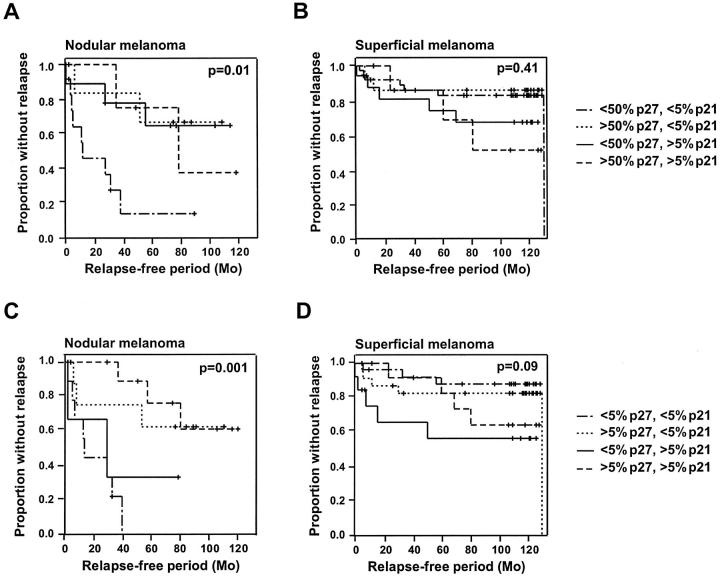
Figure 3.
Kaplan-Meier curves demonstrating the association between relapse-free survival and expression of p21WAF1/CIP1 and p27Kip1 in nodular (A and C) and superficial (B and D) melanoma. In A and B, p27Kip1 was considered as high when more than 50% of the tumor cells stained positive, and in B and D, when more than 5% expressed the protein. In all cases, p21WAF1/CIP1 was considered as high when more than 5% of the cells showed positive p27Kip1 immunoreactivity. The different combinations of high and low p21WAF1/CIP1 and p27Kip1 are indicated.

