
Figure 2.
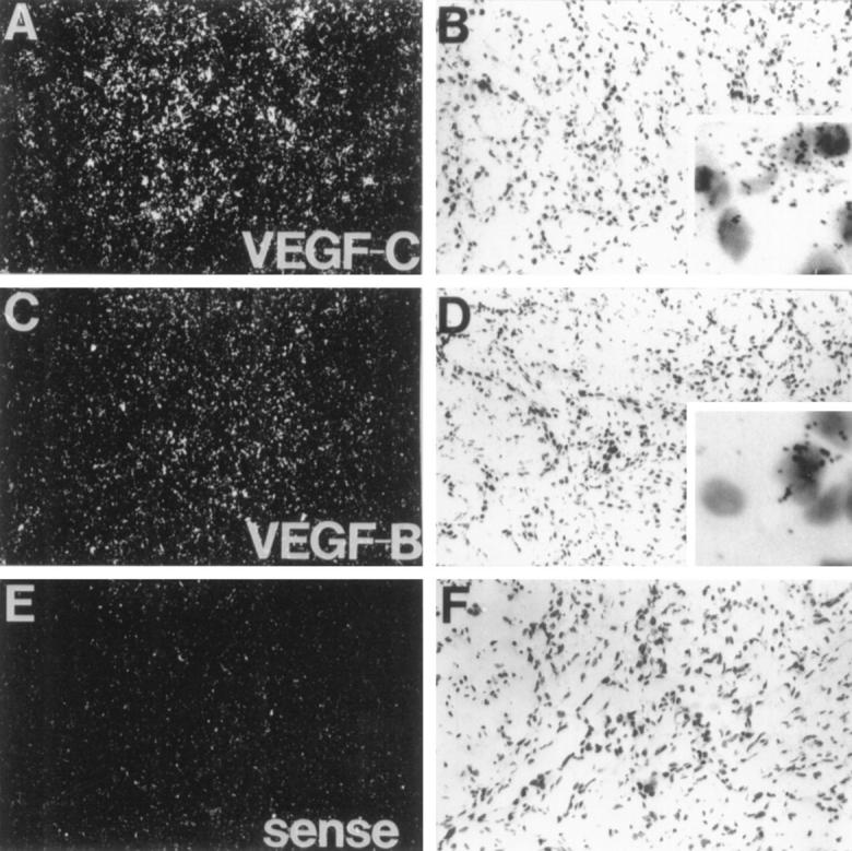
In situ hybridization analysis of VEGF-B and VEGF-C mRNAs in a head and neck carcinoma. Dark-field (A, C, and E) and light-field (B, D, and F) exposures are shown. Magnification, ×110. Insets: Higher magnification (×800). Note that the signals originate from the squamous cell carcinoma cells.